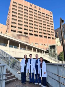
Dr. Terguunbileg Batsaikhan’s Training at Juntendo University Hospital
Primary liver cancer presents a major global public health challenge. Unfortunately, Mongolia leads the world with the highest incidence and mortality rates. In 2021, the liver cancer incidence rate in Mongolia was alarmingly high at 85.6 cases per 100,000 population, which is nine times higher than the global average and three times higher than Japan’s. HCC treatment strategies include various modalities, such as liver resection, transplantation, ablation, intravascular treatments, chemotherapy and radiotherapy. Despite advancements, challenges persist. The efficacy of anti-cancer drugs remains limited, and the scarcity of organ donors poses a significant obstacle for liver transplantation. Furthermore, not all patients with liver cancer are candidates for surgery. In response to these challenges, less invasive techniques such as ablations have become integral to clinical practice.
Ablation treatments were first introduced in Japan 40 years ago, starting with percutaneous ethanol injection therapy. Since then, the first generation of microwave ablation, radiofrequency ablation, second-generation microwave ablation technologies have been incorporated into clinical practice. Thermal ablations, including RFA and MWA, are commonly used for HCC treatment, and numerous studies are being conducted worldwide. One such study, the SURF trial from Japan, aimed to compare survival rates between patients undergoing surgery and those treated with RFA. This randomized controlled trial, conducted across 49 institutions, included 309 patients with HCC (tumor diameter ≤3 cm and ≤3 nodules) treated between 2009 and 2015. SURF trial revealed that OS and RFS were not significantly different between patients undergoing surgery and RFA for small HCC.
Image guidance methods, such as ultrasound, CT, and MRI, are used for HCC ablation treatment. Among these, ultrasound is the simplest, least expensive method and does not expose the patient to radiation. Ablation treatment can be performed with only an ultrasound device and an ablation machine equipped with an MWA antenna or RFA electrode. However, this can be considered the most basic requirement for the procedure. This baseline level is inadequate in some cases in terms of treatment safety and efficacy. Currently, the National Cancer Center of Mongolia operates at this level.
Professor Shiina and his team at Juntendo University Hospital use advanced technologies, including fusion imaging and contrast-enhanced ultrasound, along with unique techniques such as changing the patient’s position during treatment. These innovations significantly improve the precision and outcomes of ablation treatments. A dedicated ultrasound probe, co-developed by Professor Shiina and Canon, also allows for safer and more efficient procedures.
In July 2022, Professor Shiina Shuichiro and Professor Akira Sakai visited Mongolia and introduced these modern technologies to the National Cancer Center of Mongolia. At that time, I was eager to learn these techniques. In August 2023, the professors attended the HPB Surgery Week 2023 conference in Mongolia, where JIMEF offered a training opportunity at Juntendo University Hospital for a doctor from the National Cancer Center of Mongolia. I was selected for this Training and arrived in Japan in January 2024.
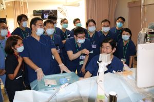
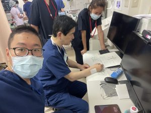
Since arriving, I have participated in and observed every ablation procedure conducted by Professor Shiina and his team. In 2024, the IVO team performed 561 ultrasound-guided percutaneous procedures at Juntendo University Hospital, including ablation, biopsies, and fiducial marker insertions. Each patient is unique, and I’ve gained valuable insights from every case—knowledge that cannot be found in textbooks or other sources. In other words, I am learning from true experts through real cases.
Table: Current Differences between the National Cancer Center of Mongolia and Juntendo University Hospital in Ablation Treatment
|
|
National Cancer center of Mongolia |
Juntendo University Hospital |
|
Anasthetics |
Local Anesthesia |
Deep sedation |
|
Image guidance method |
Ultrasound |
Ultrasound with Fusion imaging |
|
Contrast enhanced ultrasound |
– |
Available for every cases |
|
Puncturing technique |
Free hand technique |
Equipped with dedicated probe for puncturing Free hand technique available |
|
Body positioning technique/Surgery bed with mobility |
-/regular examination bed |
Available/Special designed surgery bed with mobility |
|
Artificial ascites/Artificial pleural effusion |
– |
Using these techniques more than 70% of all cases |
|
Post treatment evaluation method |
Ultrasound and laboratory results |
CECT for every cases and laboratory results |
At IVO, Juntendo University Hospital, ablation treatments are performed under deep sedation, ensuring patients experience no pain or stress and remain still during the procedure, enhancing precision and outcomes. Fusion imaging and ultrasound enable more accurate punctures and a three-dimensional view of tumors, especially in areas where standard B-mode ultrasound is ineffective. When tumors are not visible on ultrasound but detected via EOB-MRI or CECT, fusion analysis and contrast-enhanced ultrasound guide the ablation. The team also uses a specialized ultrasound probe co-developed by Professor Shiina, ensuring full visibility of the puncture path and reducing complications. A special designed surgery bed with mobility improves patient positioning, providing better access to hard-to-reach liver areas.
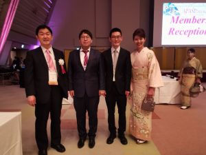
During my training in Japan, I attended two major conferences, APASL 2024 Kyoto and JRC 2024. I also had the privilege of joining the professor’s ablation training programs for both domestic and international participants.
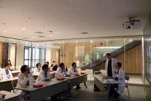
Weekly journal club meetings with the professor and trainees fostered collaboration and learning. I’m deeply grateful for the opportunity to contribute to the new ACTA guidelines.
I have also had the opportunity to participate in intravascular treatments such as transarterial chemoembolization (TACE) and hepatic arterial infusion chemotherapy (HAIC) under the guidance of Professor Hiroaki Nagamatsu. This experience has been invaluable, as HAIC new FP treatment is not yet available in Mongolia, and it is critical for managing advanced HCC cases.
Through this training, I have gained a deep understanding of how the Japanese medical system operates and how their medical staff work. I am learning a great deal from them every day.
Implementing these techniques at the National Cancer Center of Mongolia will enable us to offer minimally invasive, non-surgical treatment options to more patients. To achieve this, it is essential to acquire an ultrasound machine equipped with fusion imaging, a dedicated probe, and a special designed surgery bed with mobility. These advancements will significantly improve the quality of treatment for early-stage HCC cases in Mongolia.
I am deeply grateful to be part of this project and to have the opportunity to acquire such invaluable knowledge. On behalf of the National Cancer Center of Mongolia, as well as personally, I would like to extend my heartfelt gratitude to Professor Shuichiro Shiina, Professor Hiroaki Nagamatsu, Professor Hitoshi Maruyama, Professor Maki Tobari, and the entire Juntendo University Hospital team for their support and guidance.
I would also like to thank Professor Akira Sakai, JIMEF organization for supporting this training program. My special thanks go to Dr. Chinburen Jigjidsuren, who served as a vital bridge between the National Cancer Center of Mongolia and JIMEF. I am deeply thankful to Dr. Yumchinserchin Narangerel, head of the Department of Interventional Radiology and my mentor, as well as Dr. Erdenekhuu Nansalmaa, General Director of the National Cancer Center of Mongolia, for placing their trust in me and granting me the opportunity to study in Japan.
Finally, I would like to express my appreciation to every Japanese person who welcomed me to this wonderful country and assisted me throughout my training journey.
Dr. Terguunbileg Batsaikhan
Cooperating researcher, Juntendo University Hospital
Interventional radiologist, National Cancer Center of Mongolia
